Shared and Distinct Neural Signatures of Feature and Spatial Attention
a between-subject whole-brain machine learning approach towards the predive model of the mind
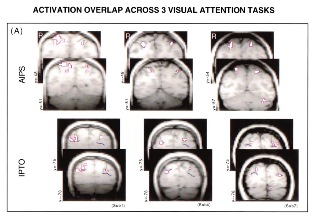
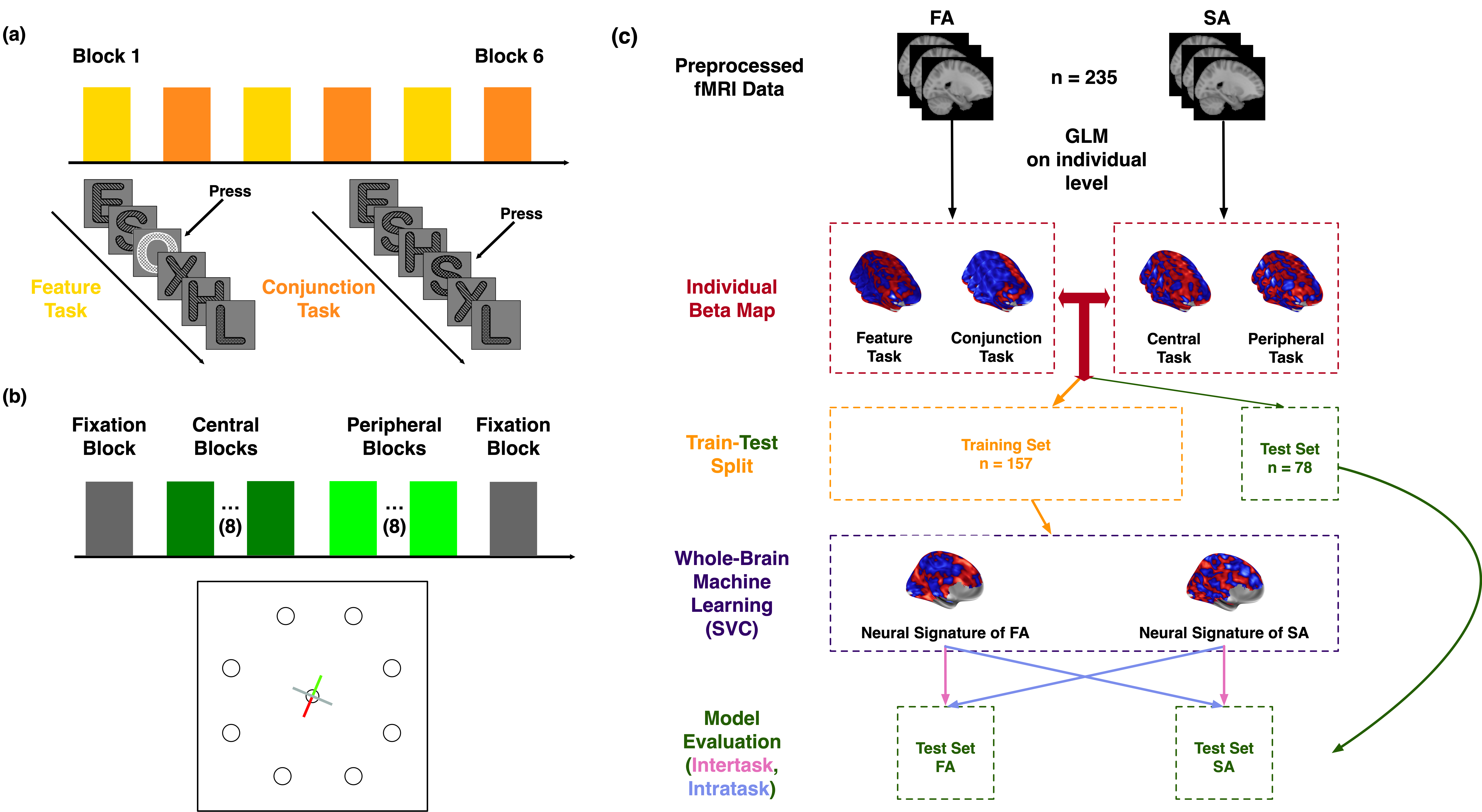
Wojciulik & Kanwisher’s research(1) suggests a common involvement of the parietal cortex in visual attention (Fig. 1). In this early research, they recorded serveral participants’ fMRI data and employed a univariate analysis. In the present study, we revisited the same paradigm (Fig. 2a, b) with two advancements:
- a larger cohort of participants (\(n=235\))
- a between-subject whole brain machine learning approach (Fig. 2c)
We replicated the same behavioral pattern (Fig. 3a) and trained two predictive models that can not only differentiate conditions in intra-task scenarios, and more interstingly, in inter-task scenarios (Fig. 3b). This suggests both shared and distinct components exit in the FAS (feature attention signature) and SAS (spatial attention signature).
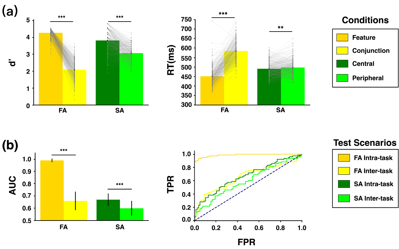
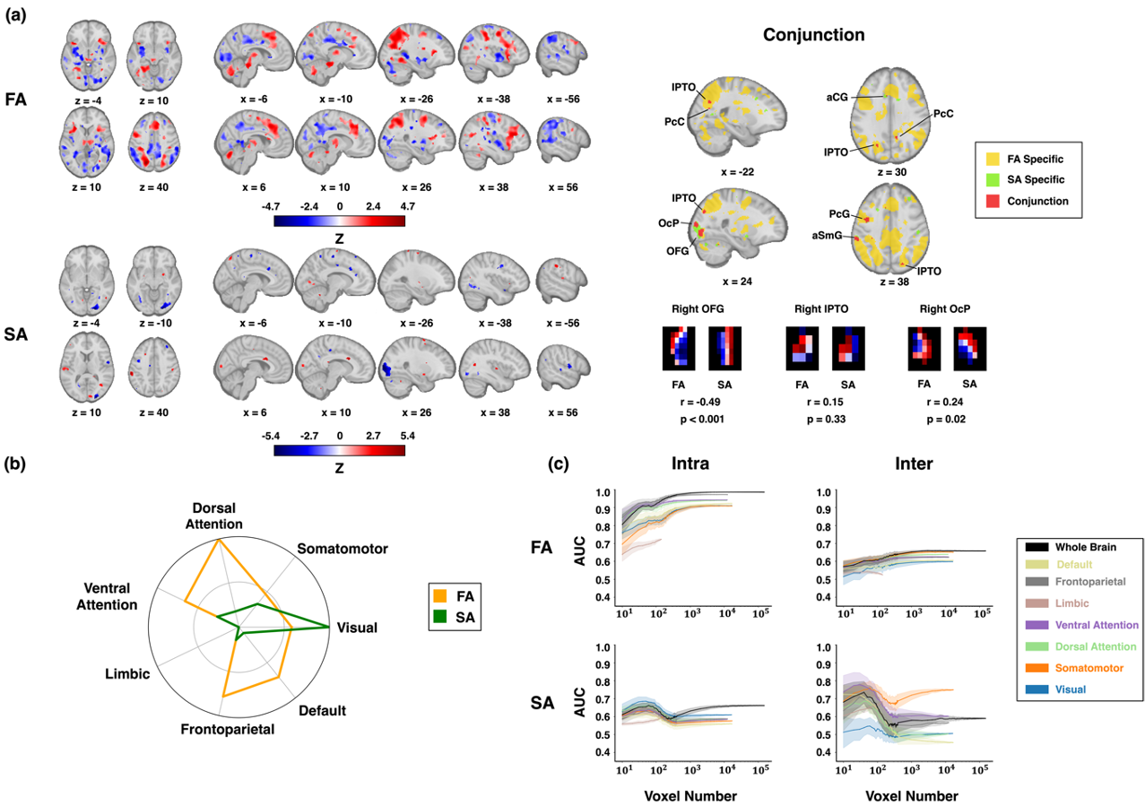
Fig. 4a shows the FAS and SAS at the whole brain level. While univariate analysis applies a boolean map with a cut-off p-value to identify the conjunction regions as the common neural subtrate, we argue that even the same region may have different pattern under different mental processes (Fig. 4a, right). When summarized at the network level, different portfolios emerge (Fig. 4b, left). More interesting, when retrained the predictive model on a network basis, the most prominent regions associated with the FAS were the dorsal and ventral attention networks, whereas the SAS was predominantly distributed within the visual network (Fig. 4c).
As we have built statistical models, we further performed the following two analyses at the cluster level:
- Single-cluster Analysis
- to test the sufficiency of one cluster
- ‘Virtual Lesion’ Analysis
- to test the necessity of one cluster
The single-cluster analysis and the “virtual lesion” analysis revealed both shared and distinct representation patterns in FA and SA (supporting table not shown), along with the engagement of a distributed brain network in processing both forms of attention (Fig. 5a).
The distributed nature is further confirmed at the voxel level (Fig.5 b).
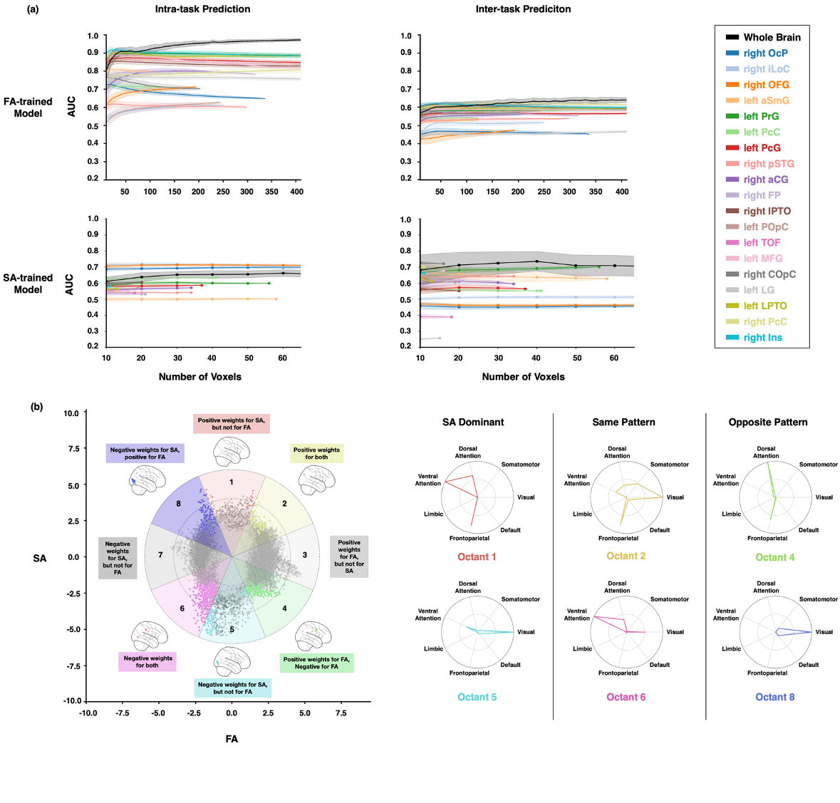
Our study challenges traditional network-centric models of attention, emphasizing distributed brain functioning in attention, and provides comprehensive evidence for shared and distinct neural mechanisms underlying FA and SA.
(1) Wojciulik, E., & Kanwisher, N. (1999). The generality of parietal involvement in visual attention. Neuron, 23(4), 747-764.
See our work at bioRxiv